Echocardiograms are one of the most common noninvasive tests performed in cardiology. Echocardiograms use ultrasound to look inside a beating heart from outside the body.
“Echocardiograms are essential for looking at the anatomy and function of a heart and monitoring how well its valves are working,” says Christine Jellis, MD, a Cleveland Clinic cardiologist. “Echocardiograms allow us to assess a person’s cardiac function with great accuracy and follow a person’s condition over time to make sure we are providing appropriate treatment.”
How Does an Echocardiogram Work?
Cardiac ultrasound technology produces images from sound waves bounced off the heart. The images can be moving or still, depending on the type of information needed.
“The beauty of ultrasound technology is that there is no radiation associated with it,” Dr. Jellis says.
The test is quiet and painless, so there is nothing to be afraid of. You can see your heart beating on the screen and watch as blood flows through it.
Types of Echocardiograms
Cardiologists use a variety of echocardiograms for different indications and different patients:
- Transthoracic Echocardiogram (TTE). TTE is the most common form of echocardiography. TTEs can be done in a cardiologist’s office or in the hospital using portable machines that are wheeled to the bedside. “TTE provides high-quality images that give us all the information we need from most people,” Dr. Jellis says.
- Transesophageal Echocardiogram (TEE). In TEE, a thin ultrasound probe is passed down the esophagus, which sits directly behind the heart. Positioning it there shortens the distance between the heart and the probe. “The resolution is outstanding when the probe is so close to the heart,” says Dr. Jellis. “TEE gives us beautiful images of the valves or masses or clots within the heart that we can’t see well with TTE.” A local anesthetic spray is used to numb your throat, and intravenous sedation is given to relax you for the test.
- Stress Echocardiogram. A stress echocardiogram uses ultrasound to assess the heart’s function when exercising. The test can unmask dysfunction that is not apparent at rest. After moving images of the heart at rest are taken with TTE, you will exercise on a treadmill or supine bike to increase your heart rate. If exercise is not possible, the heart rate is raised with intravenous medication. Once a threshold heart rate is reached, the TTE is repeated, and the before-and-after echocardiograms are compared. “Stress echo can unmask underlying cardiac dysfunction suggestive of coronary artery disease, valve disease, or other specific indications that may be apparent only when they are induced with stress,” Dr. Jellis explains.
- Doppler Echocardiogram. Doppler is an ultrasound technique used to measure blood flow and pressure differences in the heart. Its most common use is to evaluate valve function, so it is often used in people with mitral regurgitation (in which blood leaks backward into the heart’s left atrium due to a faulty mitral valve) or aortic stenosis (a narrowing of the aortic valve). Doppler measures how sound waves change in speed as they move toward you or away from you. As blood flows toward or away from the ultrasound probe, spectral Doppler produces a distinctive whooshing sound. Color Doppler color-codes the direction of flow toward or away from the echo probe. Additionally, Doppler can reveal how fast blood is flowing through a valve and any difference in pressure across the valve. “This information enables us to determine whether valvular regurgitation or stenosis is mild, moderate, or severe,” Dr. Jellis says.
- 3D/4D Echocardiogram. 3D echocardiography—sometimes called 4D echocardiography—is a new technique that provides images of the heart in three dimensions. It allows a cardiologist to assess the function of a patient’s heart and valves in more detail and to obtain the volume of the different chambers. 3D echocardiography also is used to guide transcatheter procedures like edge-to-edge repair, paravalvular leak closure, and transcatheter valve replacement. “3D TEE and 3D TTE enabled these techniques to develop,” Dr. Jellis says. “3D/4D echocardiography allows us to see the structure of a valve in greater detail than ever before. It can be used to guide the procedural cardiologist to the exact location where a prosthesis needs to be positioned. …3D information also can help us diagnose and assess the severity of the problem to determine if someone actually needs a procedure.”
- Strain Imaging. Strain imaging is a way of measuring how well a heart is functioning by assessing how much it deforms as it contracts. Comparing the shape of the heart in its relaxed state to the shape in maximal contraction provides an assessment of the degree of shape change. Strain imaging is used to asses many conditions, including chemotherapy-induced cardiac dysfunction and amyloidosis. “Strain imaging is so sensitive that it can detect reduced ventricular function before it becomes apparent by using traditional measures like ejection fraction,” Dr. Jellis says.
Why an Echocardiogram May Be Performed
Echocardiograms can be used to screen for heart problems when you have symptoms, confirm a diagnosis, assess your heart’s function, monitor a condition, or follow up on treatment. They also can be used to prepare you for heart surgery or check the outcome of your surgery.
Common symptoms that may prompt an echocardiogram are palpitations, chest pain, shortness of breath, or swelling in the ankles.
What to Expect Before, During, and After the Test
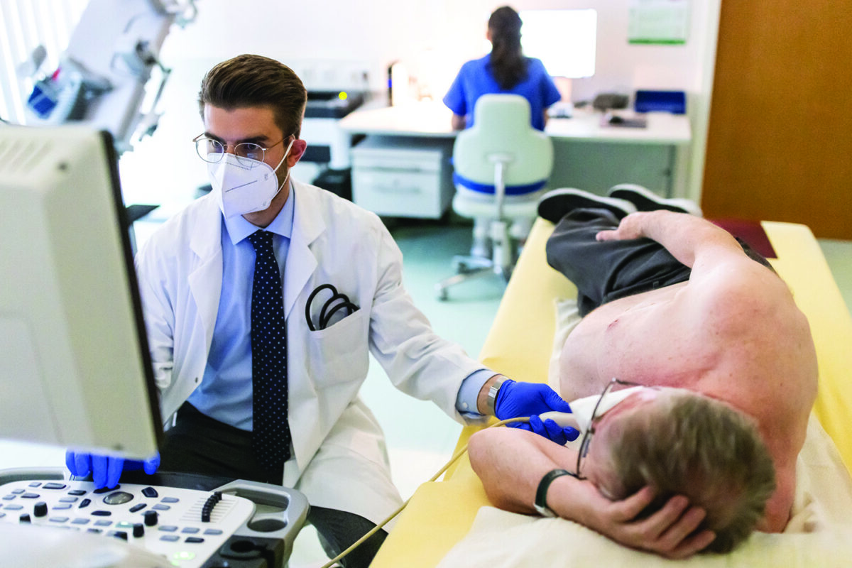
No preparation is required for a TTE. You will lie down on a bed and be asked to turn to one side. A cardiac ultrasound technician (sonographer) will attach electrodes for an electrocardiogram (ECG) to your chest. The sonographer will put a clear gel on your chest and then place an ultrasound transducer against your skin. The transducer will then be moved around to visualize specific areas of the heart while a monitor displays the images in real time. With the click of a button, the sonographer will save certain images for the cardiologist to view.
For a stress echocardiogram, the sonographer will attach ECG electrodes to your chest. A TTE will be taken. Then, you will begin exercising on the bike or treadmill. When your heart rate reaches a certain level and/or you have reached your maximal exercise capacity, another TTE will be taken for comparison. If you are receiving medication to speed up your heart rate, you will be closely monitored before and after the second TTE is taken.
Stress echocardiograms sometimes require certain medications to be withheld until the test has been completed—you’ll be notified in advance if this applies to you.
Before a TEE, you must fast for at least six hours. You will lie on a bed and be given gentle sedation for relaxation. Your mouth and throat will be sprayed with a numbing solution, or you will gargle with the solution. Then, the ultrasound probe will be passed down your esophagus. After the test is over, you will remain under supervision until you are comfortable standing and walking. You will not be allowed to drive home.
Understanding Your Echocardiogram Results
Echocardiograms are interpreted by imaging cardiologists like Dr. Jellis.
“We love the technology,” she says. “We are able to give patients and their physicians very useful information regarding their heart’s structure and function. If the function is abnormal, we can quantitate the degree of dysfunction as mild, moderate, or severe. This is incredibly useful information for guiding treatment decisions.”