A bone density scan, also called a DEXA scan/DXA scan, plays an important role in screening for osteoporosis and monitoring bone health. But does a bone density scan show cancer?
“DXA scans cannot diagnose bone cancer or bone metastases,” says Yaroslav (Slava) Gofnung, MD, a UCLA Health endocrinologist. “DXA scans are designed specifically to measure bone density to screen and diagnose osteoporosis. The DXA measures the mineral, particularly calcium, content of bone.” He notes that DXA scans can also be used to measure body composition, including body fat.
What Is a Bone Density Scan?
A bone density test is a quick, painless imaging exam that measures the amount of mineral, primarily calcium, in your bones. The results help estimate bone strength and determine whether you have normal bone density, low bone mass (osteopenia), or osteoporosis.
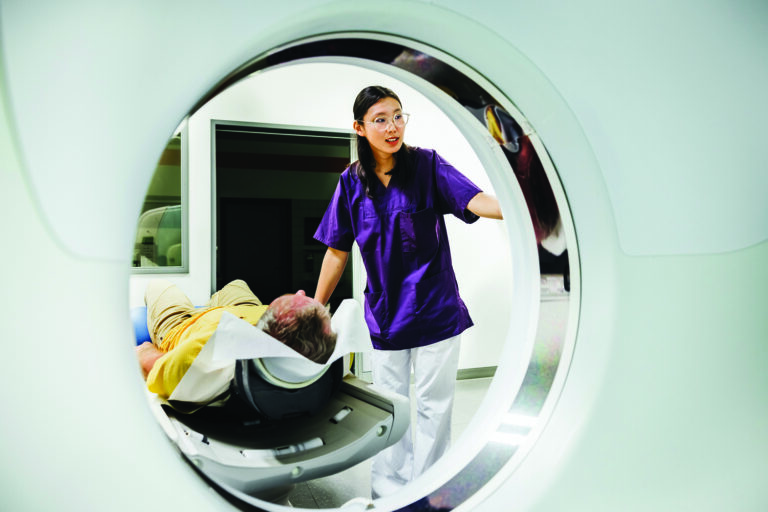
During the test, you lie on a padded table while a scanner passes over your body. Most scans focus on the hip and spine because fractures in these areas are common and can have serious consequences. The exam typically takes 10 to 20 minutes and exposes you to very little radiation, much less than a standard x-ray.
Doctors most often recommend DXA scans to:
- Screen for osteoporosis
- Estimate fracture risk
- Monitor bone loss over time
- Evaluate how well osteoporosis treatment is working
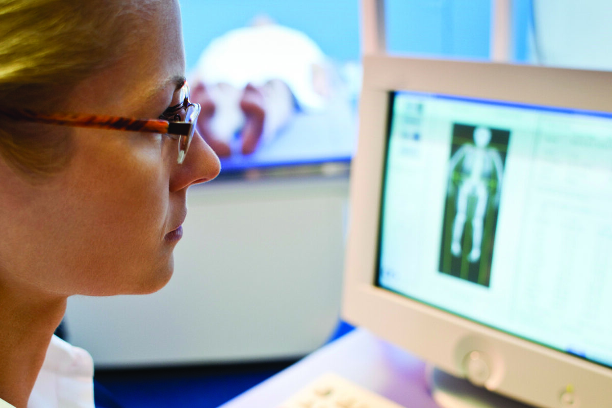
Although the scan produces images of the skeleton, its purpose is to calculate bone mineral density rather than identify tumors or other abnormalities.
Can a Bone Density Scan Detect Cancer?
No. A DEXA scan/DXA scan is not a test for bone cancer detection. It does not provide the detailed anatomical images needed to identify bone tumors or cancer that has spread to the bones.
That distinction is important because bone density and bone cancer involve two very different questions. A bone density scan asks:
- How strong are your bones?
- Are you losing bone mineral?
- Are you at increased risk for fractures?
Imaging tests for bone cancer ask different questions, such as:
- Is there a tumor inside the bone?
- Has cancer spread to the skeleton?
- Are nearby tissues involved?
A person could have completely normal bone density and still have bone cancer. Likewise, someone with osteoporosis may have very low bone density but no cancer at all.
If you have persistent bone pain, unexplained fractures, swelling, or another symptom that raises concern for cancer, your doctor will order more appropriate imaging tests rather than a DXA scan.
How Is Bone Cancer Detected?
When physicians suspect bone cancer or bone metastases, they use imaging tests that provide much more detail than a bone density test. According to Dr. Gofnung, these may include:
- X-rays, which are often the first imaging test and can identify abnormal changes in bone.
- MRI for bone cancer, which Dr. Gofnung says is “probably [the] best modality” for evaluating suspected bone tumors because it provides excellent detail of bone marrow and surrounding soft tissues.
- PET-CT or CT scans for bone cancer, which help determine the size of a tumor, whether cancer has spread, and how active the disease may be.
- Bone scans (Technetium-99 scintigraphy), which can identify areas of increased bone activity that may warrant additional evaluation.
If imaging reveals a suspicious lesion, doctors often perform a bone biopsy, removing a small tissue sample for laboratory analysis. A biopsy is the only way to definitively diagnose most bone cancers.
Bone Density Scans During Cancer Treatment
Although a bone density scan cannot detect cancer, it can play an important role in caring for people who have cancer. Some cancer treatments weaken bones, increasing the risk of osteoporosis and fractures. In these situations, doctors use DXA scans to monitor bone health before and during treatment.
Dr. Gofnung says screening bone density scans are recommended before starting, and then every two years during, several therapies known to reduce bone density, including:
- Long-term glucocorticoid (steroid) therapy
- Aromatase inhibitors, such as anastrozole, letrozole, and exemestane
- Hormone deprivation therapies, including GnRH agonists such as Lupron
- GnRH antagonists, such as Firmagon
- Radiation therapy
- Surgery that causes premature menopause by removing the ovaries
- Removal of the testicles when testosterone replacement is not possible
In these cases, the goal is not to look for cancer but to identify bone loss early so treatment can help prevent fractures.
When Should You Get a Bone Density Scan?
Most people don’t need a routine bone density test until they reach an age when osteoporosis becomes more common. Dr. Gofnung notes that, based on Endocrine Society guidelines, screening is generally recommended for all women ages 65 and older and all men age 70 and older.
You may also benefit from a bone density test if you:
- Have had a fracture after a minor fall
- Take long-term steroid medications
- Are receiving certain cancer treatments
- Have experienced early menopause
- Have a strong family history of osteoporosis
- Have other risk factors for low bone density
While a DXA scan is one of the best tools for osteoporosis diagnoses and monitoring bone health, it should not be confused with tests used to diagnose cancer.
If you have symptoms such as persistent bone pain, unexplained swelling, unexplained fractures, or concerns about cancer, talk with your healthcare provider. He or she can recommend the imaging tests that are appropriate for your situation and, if needed, arrange further evaluation.